 On the interaction of
electromagnetic fields with a human head model using computer visualization
On the interaction of
electromagnetic fields with a human head model using computer visualization
On the interaction of
electromagnetic fields with a human head model using computer visualizationAtef Z. Elsherbeni, Joseph S. Colburn, Yahya Rahmat-Samii and Clayborne D. Taylor, Jr.
With the growth of mobile communications and the close proximity of portable handsets to human beings, the interaction of radiated electromagnetic fields with nearby biological tissue is significant and an important subject of investigation. Not only is the operator's influence on the antenna gain, radiation pattern, and input impedance an issue, but there are concerns over the health effects of tissue exposed to electromagnetic energy radiated from hand-held devices. This topic of handset radiators in close proximity to human tissue has been reported previously in the literature. Different numerical techniques were applied to the problem, in addition to different head and handset radiator approximations. The finite-difference time-domain (FDTD) method is one of the more popular numerical techniques for this type of problem because prediction of electromagnetic behavior in general topologies is relatively straight forward with acceptable numerical costs.
In the following movies we place a human head phantom in close proximity to handset radiators. The main objective is to demonstrate the utility of advanced numerical electromagnetic visualization techniques for solving an important class of real-life problems. In this example a dipole antenna is used as the handset radiator and results at 900 MHz are given. The dipole antenna was fed by a voltage source with internal resistance. The head model is created from actual MRI scans which are transformed into the FDTD grid. The computational head model includes 8 different tissue types, for which the electrical properties can be prescribed at 900 MHz. For each unit cell in the discritized FDTD space, a separate conductivity, permitivity and permability can be assigned which allows for modeling very inhomogeneous dielectric materials. The procedure presented allows one to readily vary the discretization size of the computational head to meet the necessary physical resolution and/or computer resources available.
Once the head geometry is defined in the FDTD space, visualization of the computed electric and/or magnetic field values in time was considered. An animated surface plotting program was developed using the GL graphical programming language and used to view the field magnitudes and create movie files which show the time propagation of the electric and/or magnetic field through and around the head. These movie files were generated using a Sillicon Graphics workstation, and then converted to a format suitable for viewing on personal computers. Examples of these movie files are described in the Animations section.
Standards for movie software are evolving rapidly. The first four files below are QuickTime movies, converted from Dicer Animator PICS files into mov files using Apple's MoviePlayer program. Information on downloading Quicktime viewers for various platforms can be obtained from http://quicktime.apple.com/. Note that Netscape Navigator 3.0 has a built-in QuickTime plug-in viewer. For Macintosh users with an earlier version of Netscape, the MoviePlayer application can be downloaded from Apple software update site, in the Utilities directory. Don't forget to set up MoviePlayer as your helper application for Mime type/subtype "video/quicktime" for files with a "mov" extension (file type Moov). The movies will also work with the earlier SimplePlayer.
FDTD Visualization of EM Interaction with a Human Head

lrhead.mov (2.9 Mbyte) MRI scans and their corresponding low resolution (5mm cell size) discretized image for the axial cuts through the head.
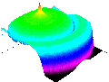
dipole.mov (4.3 Mbyte) The electric field distribution across the x-y plane at the center of a z-directed dipole antenna radiating in free space. The length of the dipole antenna is 12 cm with a 0.6 mm radius and is excited by a 900 MHz sinsoidal voltage source, with the amplitude appropriately chosen to deliver 0.6 Watts.
 hmag0.mov
(4.2 Mbyte) The magnetic field distributions across an x-y plane cut 12 cm up from the
chin. The head is excited with the field radited from a dipole antenna. The length of the
dipole antenna is 12 cm, with a 0.6 mm radius and is located approximately 6 mm away from
the head model. The dipole antenna is excited by a 900 MHz sinsoidal voltage source, with
the amplitude appropriately chosen to deliver .6 Watts. The eyes are facing the y-axis.
hmag0.mov
(4.2 Mbyte) The magnetic field distributions across an x-y plane cut 12 cm up from the
chin. The head is excited with the field radited from a dipole antenna. The length of the
dipole antenna is 12 cm, with a 0.6 mm radius and is located approximately 6 mm away from
the head model. The dipole antenna is excited by a 900 MHz sinsoidal voltage source, with
the amplitude appropriately chosen to deliver .6 Watts. The eyes are facing the y-axis.
emag0.mov
(4.3 Mbyte) The electric field distributions across an x-y plane cut 12 cm up from the
chin. The head is excited with the field radited from a dipole antenna. The length of the
dipole antenna is 12 cm, with a 0.6 mm radius and is located approximately 6 mm away from
the head model. The dipole antenna is excited by a 900 MHz sinsoidal voltage source, with
the amplitude appropriately chosen to deliver .6 Watts. The eyes are facing the y-axis.
 head.avi
(2.1 Mbyte) The three MRI scans (upper left PD weighted image, upper right T1 weighted
image, lower left T2 weighted image) and the corresponding discretized image (lower right
corner) for the axial cuts through the head.
head.avi
(2.1 Mbyte) The three MRI scans (upper left PD weighted image, upper right T1 weighted
image, lower left T2 weighted image) and the corresponding discretized image (lower right
corner) for the axial cuts through the head.